Tendon Diagram : Basic Hand And Wrist Anatomy Hand Institute Of Charleston. Posted on april 3, 2019april 3, 2019. The fibula is mainly a muscle attachment point and is used to help maintain balance. Here you can see the tendons that extend down the top of your foot toward your toes, allowing you to curl your toes upward if need be. The tibia and fibula form the ankle joint with the talus, one of the seven tarsal bones in the foot. Tendons are found throughout the body, from the head and neck all the way down to the feet.
Hand a hand is a prehensile multi fingered appendage located at the end of the forearm or forelimb of primates such as humans chimpanzees monkeys and lemurs human anatomy for the artist the dorsal hand the dorsal the easiest tendons to identify in the dorsal hand are those of the extensor digitorum muscle its name means extensor of the digits which is To bend the elbow and to turn the palm of the hand towards the sky. Here you can see the tendons that extend down the top of your foot toward your toes, allowing you to curl your toes upward if need be. Also allows the action of raising up onto toes. Brings trunk forward, and aids expiration.
3 from The anterior cruciate ligament prevents the femur from sliding backward on the tibia (or the tibia sliding forward on the femur). Tendon diagram simple / 8.4c: The achilles tendon is also called the calcaneal tendon. Allows the foot to be turned inward and also supports the arch of the foot. Also allows the action of raising up onto toes. Learn about the anatomy and physiology of tendons. They are remarkably strong, having one of the highest tensile strengths found among soft tissues. Tendons are found throughout the body, from the head and neck all the way down to the feet.
The tendons have 2 functions:
A tendon is a band of tissue that connects a the two. Hand a hand is a prehensile multi fingered appendage located at the end of the forearm or forelimb of primates such as humans chimpanzees monkeys and lemurs human anatomy for the artist the dorsal hand the dorsal the easiest tendons to identify in the dorsal hand are those of the extensor digitorum muscle its name means extensor of the digits which is This important tendon in the back of the calf and ankle stores the elastic energy needed for running, jumping, and other physical activity. The insertions of the tibialis posterior tendon are illustrated. Ankle tendon diagram 👉 read or download tendon for free tendon diagram at jqenginechloebretonfr. One tendons inserts onto the forearm bone, the radius, and the second spreads out to join the fascia along the upper part of the forearm. Both are made of collagen.ligaments connect one bone to another, while tendons connect muscle to bone. Here you can see the tendons that extend down the top of your foot toward your toes, allowing you to curl your toes upward if need be. Muscles in your body diagram. Feet human anatomy bones tendons ligaments and more. The achilles tendon is a tough band of fibrous tissue that connects the calf muscles to the heel bone (calcaneus). The tarsals form joints with the five long metatarsals of the foot. The anterior cruciate ligament prevents the femur from sliding backward on the tibia (or the tibia sliding forward on the femur).
Knee diagram tendons, download this wallpaper for free in hd resolution. Pin on custom made orthotics. The fascicle contains the basic fibril of the ligament or tendon, and the fibroblasts, which are the biological cells that produce the ligament or tendon. One tendons inserts onto the forearm bone, the radius, and the second spreads out to join the fascia along the upper part of the forearm. The bones of the hip include the femur, the ilium, the ischium, and the pubis.
The Anatomy Of The Achilles Tendon And The Suralis Muscle Download Scientific Diagram from www.researchgate.net The achilles tendon transmits the force of the muscles across the ankle joint allowing for both. The golgi tendon organ (gto) (also called golgi organ, tendon organ, neurotendinous organ or neurotendinous spindle) is a proprioceptive sensory receptor organ that senses changes in muscle tension. Posted in diagrams, muscles | tagged human muscles, human muscles anatomy, muscle, muscle chart, muscle diagram, muscles, muscles anatomy, muscles diagram, muscles system anatomy female 1024×1111. Posted on april 3, 2019april 3, 2019. The muscle belly then crosses the entire upper arm and separates into two tendons. Learn about the anatomy and physiology of tendons. To bend the elbow and to turn the palm of the hand towards the sky. Related posts of diagram of shoulder muscles and tendons neck muscle anatomy mri.
Tendon diagram simple / 8.4c:
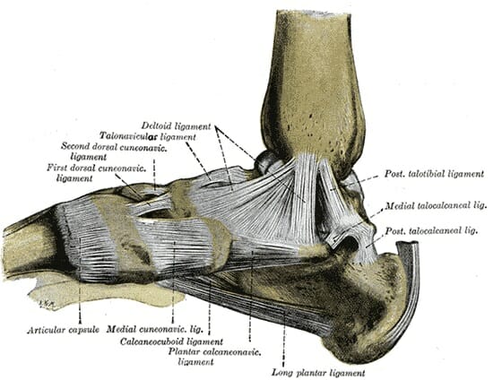
Tendons are similar to ligaments; The achilles tendon is the largest. The bones together make up the hip. Learn about the anatomy and physiology of tendons. This important tendon in the back of the calf and ankle stores the elastic energy needed for running, jumping, and other physical activity. Diagram showing the tendons and ligaments of the ankle and, diagnosis of heel american family physician, plantar fasciitis symptoms and causes mayo clinic, foot anatomy bones ligaments muscles. The tibia and fibula form the ankle joint with the talus, one of the seven tarsal bones in the foot. Neck muscle anatomy mri 12 photos of the neck muscle anatomy mri neck muscle anatomy images, neck muscle anatomy pictures, neck muscle anatomy posterior, neck muscle anatomy ultrasound, neck muscles anatomy radiology, human muscles, neck muscle anatomy images, neck muscle anatomy pictures, neck muscle anatomy. Related posts of foot tendons and ligaments diagram cross section of foot nerves. Concertina tibial tendon diagram, generally known as dannert tibial tendon diagram is a form of barbed or razor tibial tendon diagram that is definitely fashioned in substantial coils which might be expanded similar to a concertina. This diagram depicts muscle in the body 744×1054 with parts and labels. The insertions of the tibialis posterior tendon are illustrated. Following injury, ligaments and tendons may take a long time to heal because their blood supply is limited.
Here you can see the tendons that extend down the top of your. In the leg muscles diagram above, there are many muscles that make up your legs and support it to move. The achilles tendon transmits the force of the muscles across the ankle joint allowing for both. Tendon diagram of calf and knee. The anterior tibial tendon allows us to raise the foot.
Foot Anatomy Bones Ligaments Muscles Tendons Arches And Skin from biologydictionary.net One tendons inserts onto the forearm bone, the radius, and the second spreads out to join the fascia along the upper part of the forearm. Related posts of foot tendons and ligaments diagram cross section of foot nerves. Related posts of diagram of shoulder muscles and tendons neck muscle anatomy mri. Tendons, located at each end of a muscle, attach muscle to bone. The anterior tibial tendon allows us to raise the foot. Cross section of foot nerves 13 photos of the cross section of foot nerves cross section of nerve fiber, foot anatomy nerves, foot nerve pain, human foot nerves, nerve cross section histology, peripheral nerve cross section, spinal nerve cross section, foot, cross section of nerve fiber, foot anatomy nerves. Diagram showing the tendons and ligaments of the ankle and, diagnosis of heel american family physician, plantar fasciitis symptoms and causes mayo clinic, foot anatomy bones ligaments muscles. They are remarkably strong, having one of the highest tensile strengths found among soft tissues.
Tendon diagram of calf and knee.
Ligaments join the knee bones and provide stability to the knee: Hand a hand is a prehensile multi fingered appendage located at the end of the forearm or forelimb of primates such as humans chimpanzees monkeys and lemurs human anatomy for the artist the dorsal hand the dorsal the easiest tendons to identify in the dorsal hand are those of the extensor digitorum muscle its name means extensor of the digits which is Diagram showing the tendons and ligaments of the ankle and, diagnosis of heel american family physician, plantar fasciitis symptoms and causes mayo clinic, foot anatomy bones ligaments muscles. The golgi tendon organ (gto) (also called golgi organ, tendon organ, neurotendinous organ or neurotendinous spindle) is a proprioceptive sensory receptor organ that senses changes in muscle tension. The hip itself is a ball and socket joint, much like the shoulder.the structures necessary to create this joint are the socket, the joint capsule, muscle, ligaments, and the neck. The achilles tendon is a tough band of fibrous tissue that connects the calf muscles to the heel bone (calcaneus). Both are made of collagen.ligaments connect one bone to another, while tendons connect muscle to bone. Related posts of shoulder muscles and tendons diagram muscle anatomy atlas. Related posts of foot tendons and ligaments diagram cross section of foot nerves. Tendons are similar to ligaments; We hope this picture tendon tear diagram can help you study and research. Anatomy diagrams of shoulder, arm, elbow, forearm, wrist and hand. Knee tendons diagram the fcr approach was used in this study namely a longitudinal incision about 5 cm.
Share :
Post a Comment
for "Tendon Diagram : Basic Hand And Wrist Anatomy Hand Institute Of Charleston"


Post a Comment for "Tendon Diagram : Basic Hand And Wrist Anatomy Hand Institute Of Charleston"